
 中文
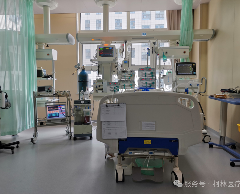
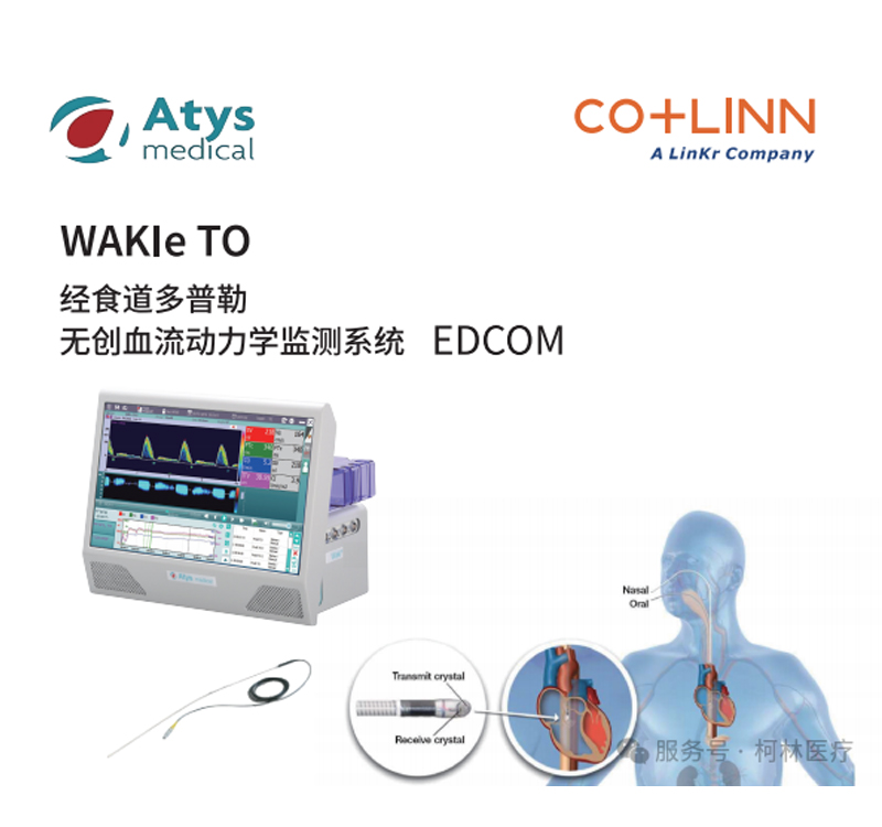
中文In the Emergency Intensive Care Unit (EICU), every second matters for a patient's survival. For patients with hemodynamic instability, how to quickly and accurately obtain circulatory data has always been the core and challenge of clinical decision-making. Faced with traditional monitoring methods, clinicians often have to weigh the "invasive risk" against the "accuracy of data." Recently, a hospital EICU introduced a transesophageal Doppler non-invasive hemodynamic monitoring system (Registration Number: Guo Xie Zhu Jin 20153061068). Through preliminary application on three patients in different conditions, it has broken the department's original concerns about this technology and provided a new perspective for the circulatory management of critically ill patients.
Breaking Concerns: From "Cumbersome" to "Standardized" Experience Transformation
Before implementing this technology, the EICU medical team generally had the following concerns: Is the insertion of the Doppler probe cumbersome? Is it difficult to insert the probe when the patient is conscious? Is the accuracy of the monitoring results reliable? However, after bedside application on several patients with different states of consciousness and conditions, these concerns were gradually eliminated. The ease of operation, the accuracy of the monitoring results, and patient tolerance were all consistently recognized by frontline clinical staff.
Clinical Case Focus: When Atrial Fibrillation Meets Cerebral Infarction, Who Controls Blood Flow?
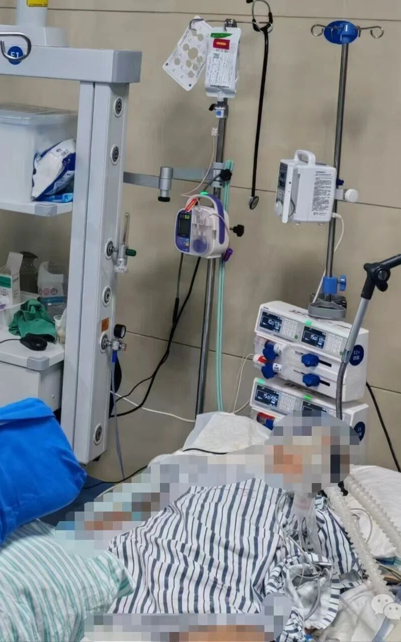
To more intuitively present the clinical value of this technology, a typical case was selected for analysis. The patient was a 65-year-old male admitted for "ischemic stroke," with a history of "persistent rapid atrial fibrillation." During EICU treatment, precise assessment of his cardiac function and volume status became crucial for treatment.
1. Limitations of Traditional Assessment Methods
Although the patient had persistent atrial fibrillation, there was no organic heart disease, so his left ventricular ejection fraction (EF) and brain natriuretic peptide (BNP) were within normal ranges. However, clinical experience indicates that rapid atrial fibrillation affects hemodynamics far beyond what these two indicators can cover.
2. The "Breakthrough" of Doppler Transesophageal Probe
At the bedside, the critical care staff successfully inserted the Doppler transesophageal probe into the patient's esophagus via the mouth in just 3 minutes, immediately obtaining typical monitoring values and clear monitoring images.
3. Intuitive Pathophysiological PresentationReal-time monitoring images clearly show that during persistent atrial fibrillation, the atria are in a state of ineffective quivering and fail to actively pump blood into the ventricles, leading to a reduction in end-diastolic volume. According to the classic Frank-Starling mechanism, the shortened initial length of myocardial fibers results in weakened ventricular contractility. This impact is more pronounced in patients with rapid ventricular rates; the significantly shortened diastole means the ventricles (especially the left ventricle) begin the next contraction before being fully filled, creating a "deficit" cycle that lowers cardiac output.
4. Alignment of Data and Clinical FindingsThe transesophageal Doppler system promptly captured these subtle changes—insufficient left ventricular ejection function while other myocardial contractility parameters remained normal. This result is highly consistent with parameters from other monitoring devices and aligns with echocardiography and BNP results, providing a solid basis for clinical decision-making.
5. Technical Advantages: The "Optimal Balance" between Invasive and Non-invasiveThree core advantages were fully verified in the EICU:
- Decision-making Cornerstone: Precise monitoring values accurately reflect left heart function changes, especially in arrhythmias like AFib, revealing the core hemodynamic shifts.
- Efficiency: Fast and user-friendly placement. For trained critical care professionals, the probe can be positioned and monitoring started within 3 minutes, minimizing interference with emergency workflows.
- Patient Benefit: High tolerance and cooperation. The probe design ensures minimal discomfort, with patient compliance exceeding expectations even without deep sedation.
Within the technological framework of hemodynamic monitoring, the transesophageal Doppler system demonstrates a unique strategic positioning. Compared to invasive pulmonary artery catheterization (PAC), it avoids the associated trauma, risks, and operational complexity; compared to bioimpedance methods, it overcomes the issues of external interference and data inaccuracy.
Transesophageal Doppler non-invasive hemodynamic monitoring is not intended to replace existing technologies. Instead, it offers clinicians a new alternative characterized by beingnon-invasive, safe, precise, and convenient, providing fresh guidance for circulatory management in critical care, emergency medicine, and perioperative settings.